

_01.jpg)
_02.jpg)
Silver nanocolloids have great antimicrobial effects. But what is the potential risk for toxicity?
August 7, 2012
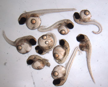
This photo, published in the journal Environmental Science
& Technology, shows malformations of medaka fish
embryos when exposed to silver nanocolloids,
whose antimicrobial effects are valued for health care products.
A study by researchers at the Arnold School of Public Health has found that nano-size silver particles dispersed in liquid – also known as silver nanocolloids (SNC) – appear to disrupt the process by which vertebrate embryos form and develop.
The team of international researchers, led by Dr. Shosaku Kashiwada, a post-doctoral fellow at the Arnold School’s Department of Environmental Health Sciences at the time of study, and Dr. Tom Chandler, Dean of the Arnold School of Public Health, examined the toxic effects of SNC on medaka fish. The results were published recently in Environmental Science and Technology, a journal of the American Chemical Society.
Morphological changes that occurred in medaka fish embryos included cardiovascular malformations, ischemia, underdeveloped eyes and central nervous system, and kyphosis (curvature of the spine). The changes to the fish embryos appear to be caused by the disruption of the expression of specific genes key to normal vertebrate development.
The study is timely. High-tech industries and medicine are increasing their use of nanotechnology, which is expected to have a $1.5 trillion global market by 2015. While “such technology offers numerous benefits, there are mounting concerns regarding the potential environmental and human health risks associated with exposure to nanomaterials,” said the researchers.
In particular, silver nanomaterials, including nanocolloids and nanoparticles, are the most widely used because of their disinfectant properties and are the major components of healthcare products. However, silver nanomaterials also have a greater opportunity to affect aquatic environments through emissions from municipal and other wastewater facilities.
The researchers found that SNC were capable of crossing the protective medaka egg membrane and reaching the embryonic mass.
Among the study’s conclusions:
- SNC impact on medaka embryos was dependent on the stage of development at exposure. For example, SNC exposure at stage 21 (one day and 10 hours) impacted brain and neural fold formation.
- SNC crossed the egg chorion and resulted in acute toxicity and adverse malformations in the embryos, including cardiovascular diseases, short tail development with kyphosis, and deformities of the brain, eyes and spine.
- In addition to problems associated with the cardiovascular system, SNC caused gross malformations in central nervous system development in medaka embryos.
Because very little is known about the specific mechanisms and modes of action regarding the toxicity of silver nanoparticles, more research on silver toxicity is needed; particularly with the increased use of such nanomaterials in consumer products and the mounting concerns over the potential environmental and human health risks associated with exposure to nanomaterials, the researchers said.
Other Arnold School researchers involved in the study included Dr. Maria Ariza, Dr. Tomohiro Kawaguchi, Dr. Sumith Jayasinghe, and Karin Gartner. The study included researchers from USC’s Nano Center and Toyo University in Japan.